BFMS-L_UFGJ_1000_900_458_D
The Basic Fluorescence Microscopy System – Deep Brain (150 µm to 8 mm depth) contains all the items necessary to do deep-brain calcium imaging of freely-moving animals.
→ 상품 상세 정보 : Basic Fluorescence Microscopy System – Deep Brain
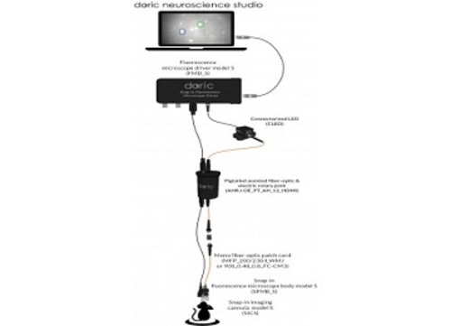
The Basic Fluorescence Microscopy System – Deep Brain (150 µm to 8 mm depth) contains all the items necessary to do deep-brain calcium imaging of freely-moving animals. This system includes specifically:
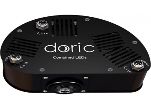
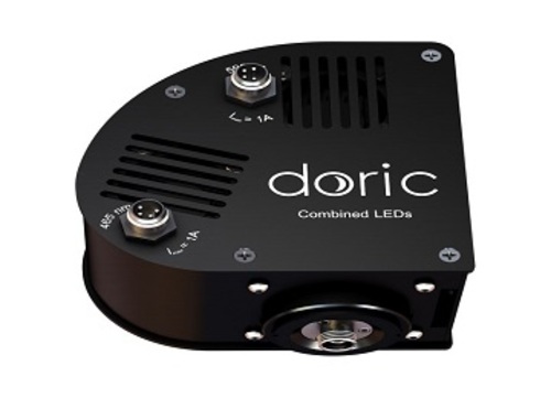
– Connectorized LED or Ce:YAG Optical Head
– Fluorescence Microscope Driver Model L
– Snap-in Fluorescence Microscope Body Model L
– Snap-in Imaging Cannula Model L (3x)
– Protrusion Adjustment Ring Set Model L
– Pigtailed Assisted Fiber-optic & Electrical Rotary Joint
– Fluorescence Microscope Holder
– Clamp for Fluorescence Microscope Holder
– Fluorescence Microscope Snapping Tool
– Dummy Microscope
– Doric Neuroscience Studio for control and analysis
– All electrical cables and optical patch cords
Notes:
• The ultralight fiberglass jacket is lighter and more flexible, the lightweight metal jacket is more robust but heavier.
• The standard electrical cable length is 1000 mm. Other values on request (up to 3000 mm).
• The optical fiber length is adjusted to fit the desired electrical cable length.
The Basic Snap-in Fluorescence Microscope Body is offered in two models: S or L. Both models have the dichroic beam-splitter, M3 optical connector, CMOS sensor etc. Each CMOS has a serial number stored within its cable that points to a specific set of mask correction filters recognizable to our software package. Model L has a 0.5 NA objective lens within its body while Model S has a plan-parallel plate instead and relies on the objective lens within the model S imaging cannula to create an image on the CMOS. When used for deep brain imaging, the fluorescence microscope body is used with an implantable imaging cannula that transfers the image from its bottom to its top.
The next figure shows the fluorescence intensity traces of 16 cells expressing GCaMP6 in the D2 receptor-expressing medium spiny neurons in the indirect pathway of striatum. The movie was acquired in the striatum (about 4 mm deep) in a freely-moving mouse. Courtesy of Dr. Akiyo Natsubori (experimenter), and Dr. Kenji Tanaka (PI) from the Department of Neuropsychiatry, School of Medicine, Keio University.


