OSMS-S_UFGJ_1000_900_458/604
The OSFM systems include the Ce:YAG & Blue Fiber Light Sources in order to synchronize the fluorophore excitation light with the opsin activation light output in the same optical fiber. This system contains all the items necessary to do surface brain calcium imaging (<150 µm depth) synchronized with opsin activation of freely-moving animals.
→ 상품 상세 정보 : Optogenetically Synchronized Fluorescence Microscope System – Surface
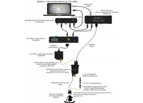
This system contains all the items necessary to do surface brain calcium imaging (<150 µm depth) synchronized with opsin activation of freely-moving animals. The OSFM Fluorescence Microscopy System – Surface includes specifically:
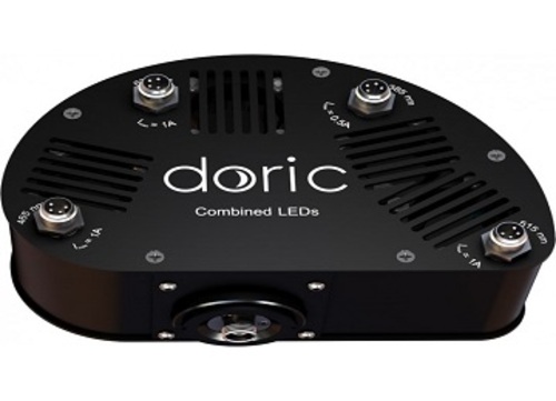
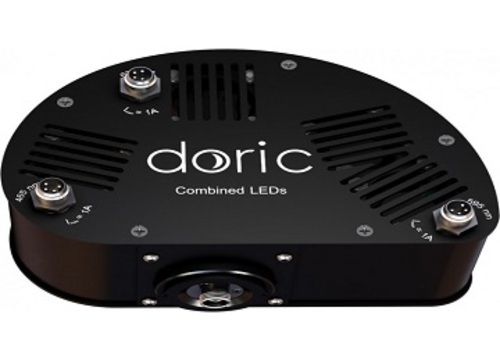
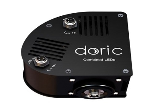
– Ce:YAG + LED (465 nm) or Laser (450 nm) Optical Head
– Ce:YAG + LED (465 nm) or Laser (450 nm) Driver
– Optogenetics TTL Generator 4-channel
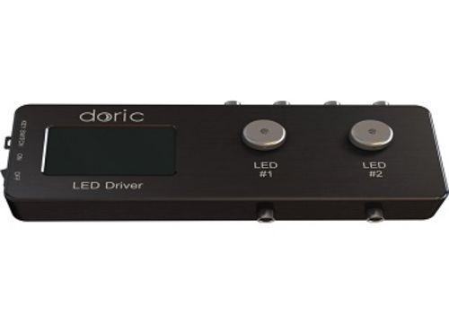
– Fluorescence Microscope Driver Model S
– OSFM Microscope Body Model S
– Snap-in Imaging Cannula Model S (3x)
– Protrusion Adjustment Ring Set Model S
– Pigtailed Assisted Fiber-optic & Electrical Rotary Joint
– Fluorescence Microscope Holder
– Clamp for Fluorescence Microscope Holder
– Fluorescence Microscope Snapping Tool
– Dummy Microscope
– Doric Neuroscience Studio for control and analysis
– All electrical cables and optical patch cords
The Optogenetically Synchronized Fluorescence Microscope or OSFM, combines fluorescence imaging and optogenetic stimulation/inhibition capabilities within the miniature fluorescence microscope. It can be used for freely-moving or head-fixed configurations. To avoid cross talk between optogenetic stimulation and fluorescence imaging, the OSFM hardware provides for at least two distinct spectral bands for light activation or fluorophore excitation (like blue and yellow) and at least two distinct spectral bands for imaging of fluorophores (like green and red). Either channel, blue-green or yellow-red can be used for opsin activation/inhibition or for calcium indicator excitation and imaging. As the field of opsins and calcium indicators is very dynamic, those spectral bands can be tailored to specs. For now, GCaMP6 + NpHR3.0 and RCaMP2 + ChR2 microscope versions are available.
Notes:
• The ultralight fiberglass jacket is lighter and more flexible, the lightweight metal jacket is more robust but heavier.
• The standard electrical cable length is 1000 mm. Other values on request (up to 3000 mm).
• The optical fiber length is adjusted to fit the desired electrical cable length.


