OSFM_S_UFGJ_1000_458/604
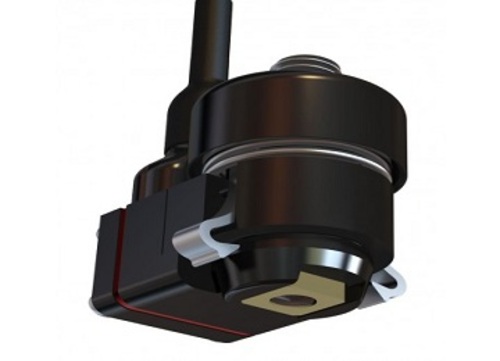
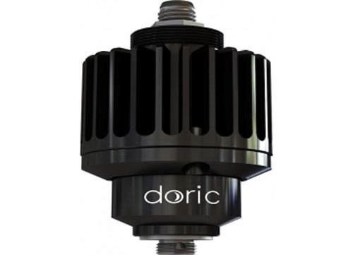
The Optogenetically Synchronized Fluorescence Microscope or OSFM, combines fluorescence imaging and optogenetic stimulation/inhibition capabilities within the miniature fluorescence microscope.
→ 상품 상세 정보 : Optogenetically Synchronized Fluorescence Microscope Bodies
The Optogenetically Synchronized Fluorescence Microscope or OSFM, combines fluorescence imaging and optogenetic stimulation/inhibition capabilities within the miniature fluorescence microscope. It can be used for freely-moving or head-fixed configurations. To avoid cross talk between optogenetic stimulation and fluorescence imaging, the OSFM hardware provides for at least two distinct spectral bands for light activation or fluorophore excitation (like blue and yellow) and at least two distinct spectral bands for imaging of fluorophores (like green and red). Either channel, blue-green or yellow-red can be used for opsin activation/inhibition or for calcium indicator excitation and imaging. As the field of opsins and calcium indicators is very dynamic, those spectral bands can be tailored to specs. For now, GCaMP6 + NpHR3.0 and RCaMP2 + ChR2 microscope versions are available.
Notes:
• The ultralight fiberglass jacket is lighter and more flexible, the lightweight metal jacket is more robust but heavier.
• The standard electrical cable length is 1000 mm. Other values on request (up to 3000 mm).
• The optical fiber length is adjusted to fit the desired electrical cable length.
• Every microscope body comes with a protective cap.
Under is the ΔF/F0 analysis of the video. The image shows 25 traces (ΔF/F0 up to 10%) of the >50 cells found in the movie.


